Spectrum and Color Results
The same coating can appear different under another source or viewing angle even when its materials and thicknesses do not change. The Spectrum pages show how the structure reshapes the incident spectrum, while the Color pages convert that spectrum into chromaticity, display color, and color differences.
Example Setup
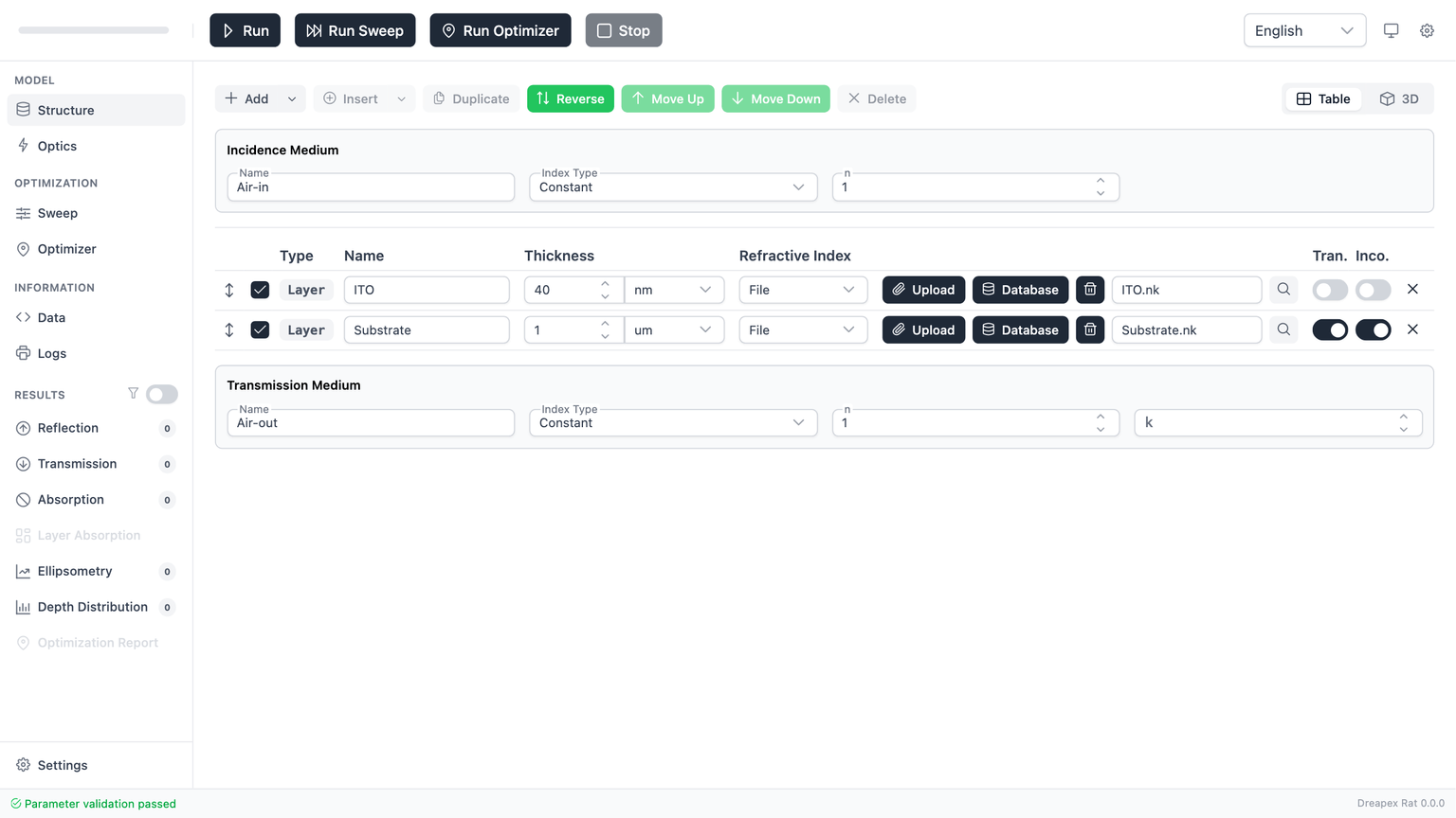
This page uses a single-layer TiO₂ interference coating to demonstrate reflected color under D65 and its change with incidence angle. Light enters from the top air side. The BK7 substrate is a 1 mm incoherent transparent layer, followed by air as the bottom medium.
Structure direction: top medium → bottom medium
| Object | Configuration |
|---|---|
| Top medium | Air, n = 1 |
| TiO₂ interference layer | 100 nm, file-based refractive index |
| BK7 substrate | 1 mm, file-based refractive index, Transparent, Incoherent |
| Bottom medium | Air, n = 1 |
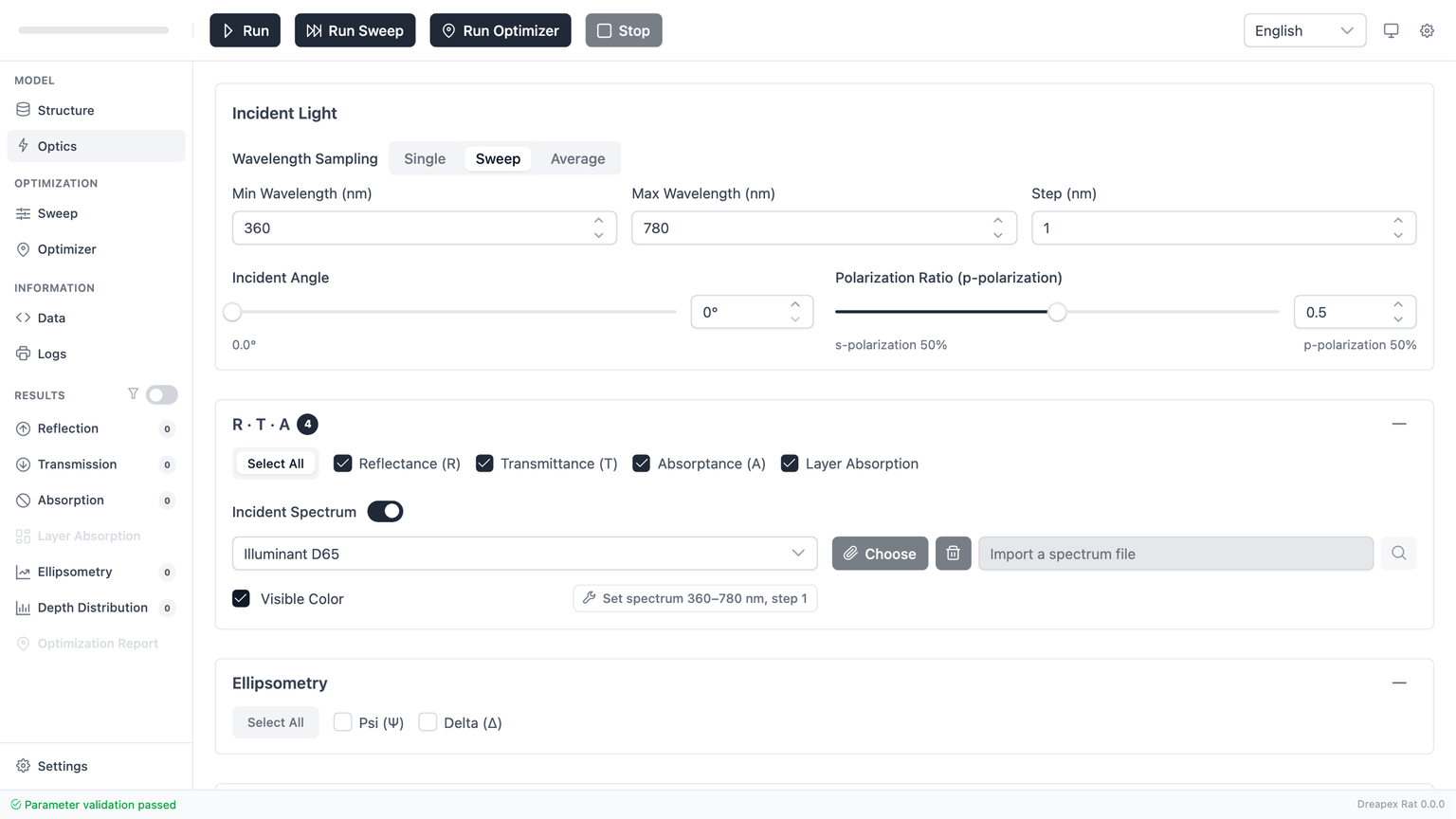
Optics uses unpolarized normal incidence with R, T, A, Incident Spectrum, and Visible Color enabled. Color integration uses the relative D65 spectral power distribution and the 2° observer.
| Parameter | Value |
|---|---|
| Wavelength mode | Sweep |
| Wavelength range | 360–780 nm |
| Step | 1 nm |
| Incidence angle | 0° |
| p-polarization ratio | 0.5 (unpolarized) |
| Incident spectrum | Illuminant D65 |
| Visible Color | Enabled |
| Observer | 2° |
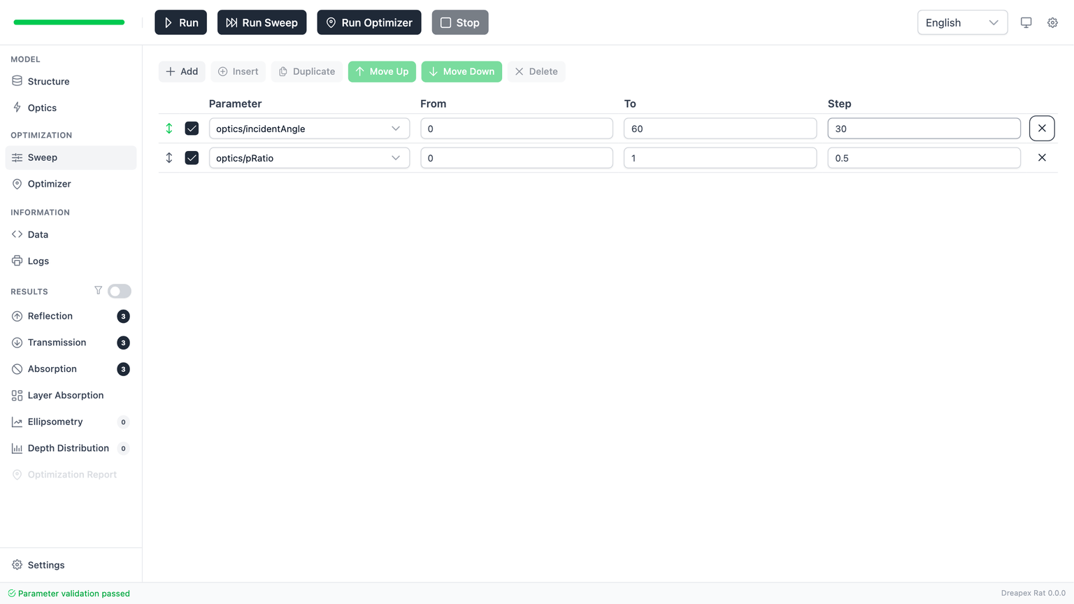
The angle sweep changes only incidence angle and uses 0°, 30°, and 60°. This isolates angular color shift under the same structure and source.
Generation Requirements
Reflection / Transmission / Absorption Spectrum requires a wavelength-resolved result, the corresponding R / T / A detector, and an enabled incident spectrum.
Reflection / Transmission / Absorption Color also requires all of the following:
- Enable
Incident Spectrum. - Enable
Visible Color. - Set the wavelength range exactly to
360–780 nm. - Set the wavelength step exactly to
1 nm. - Enable the
R,T, orAdetector you want to analyze.
The 360–780 nm / 1 nm grid is a Dreapex TMM generation requirement, not a claim of measurement conformity with ISO/CIE 11664-3:2019.
Set spectrum 360–780 nm, step 1 applies the required wavelength settings and enables color calculation in one action. Rerun after changing the source, observer, or sampling. A result keeps the observer and white point used during its run and does not change with later settings.
A custom File spectrum is read as relative spectral power per nanometre; its overall amplitude may be scaled arbitrarily. Convert photon-count or per-frequency spectra to power per nanometre first, or the color weighting will be systematically shifted.
Spectrum Results
Reflection Spectrum, Transmission Spectrum, and Absorption Spectrum combine the current incident spectrum with the structure's R / T / A response. Each plot shows both the relative source spectrum and the reflected, transmitted, or absorbed spectral intensity.
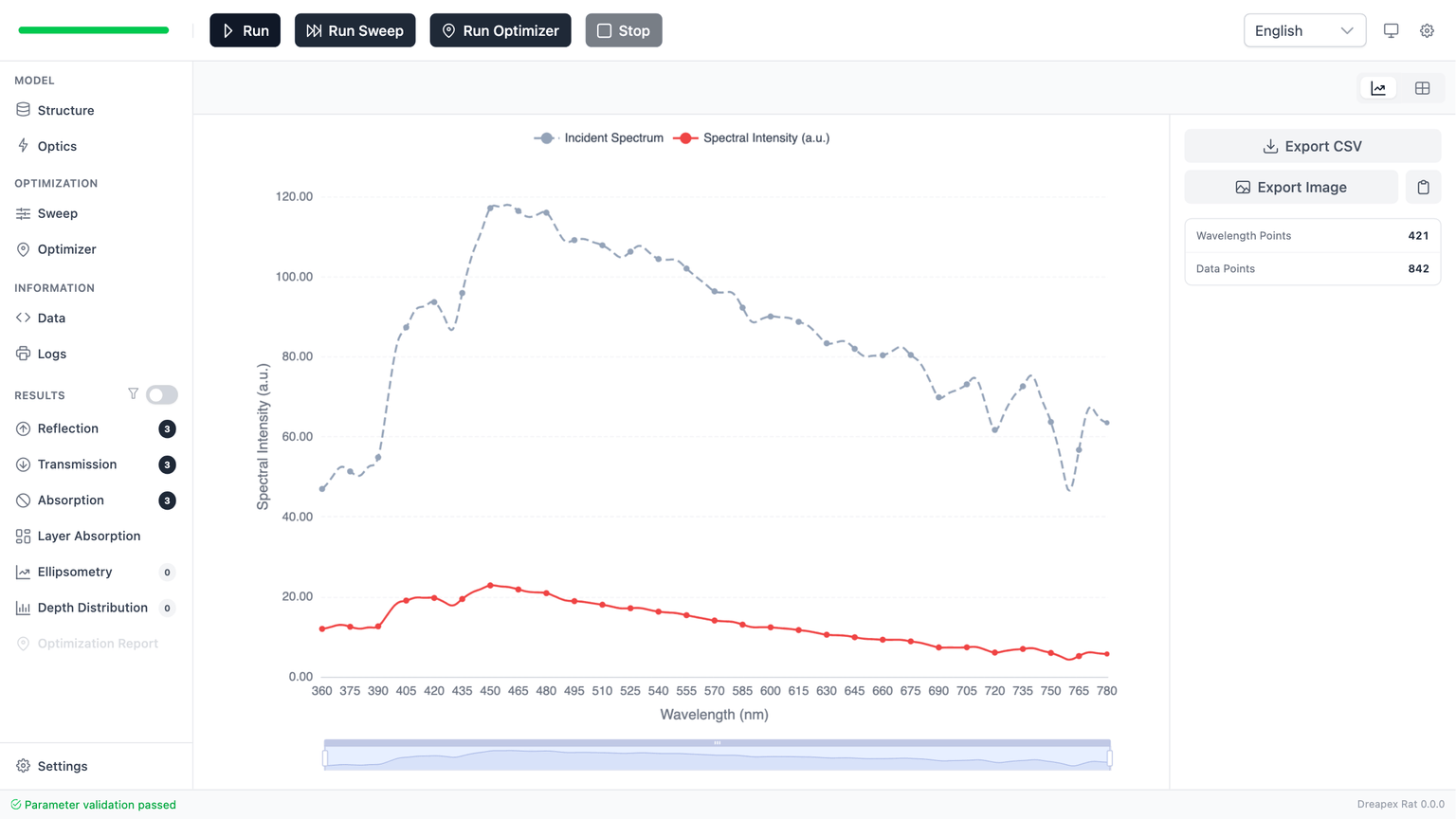
Raw R / T / A describes the structure's response at each wavelength. The weighted spectrum shows where the specified source actually supplies energy. Changing the source changes the weighted spectrum and color result, but it does not change raw R / T / A for the same structure and optical conditions.
Chromaticity Diagram
The Color page opens with the chromaticity diagram for the observer frozen into the result; its coordinates and terminology follow CIE 015:2018, Colorimetry, 4th Edition. The 2° observer uses the CIE 1931 xy diagram, with optional gamut triangles, white points, Planckian locus, and wavelength labels. The 10° observer uses the CIE 1964 locus; gamut triangles and preset white points are hidden there to avoid mixing coordinate systems.
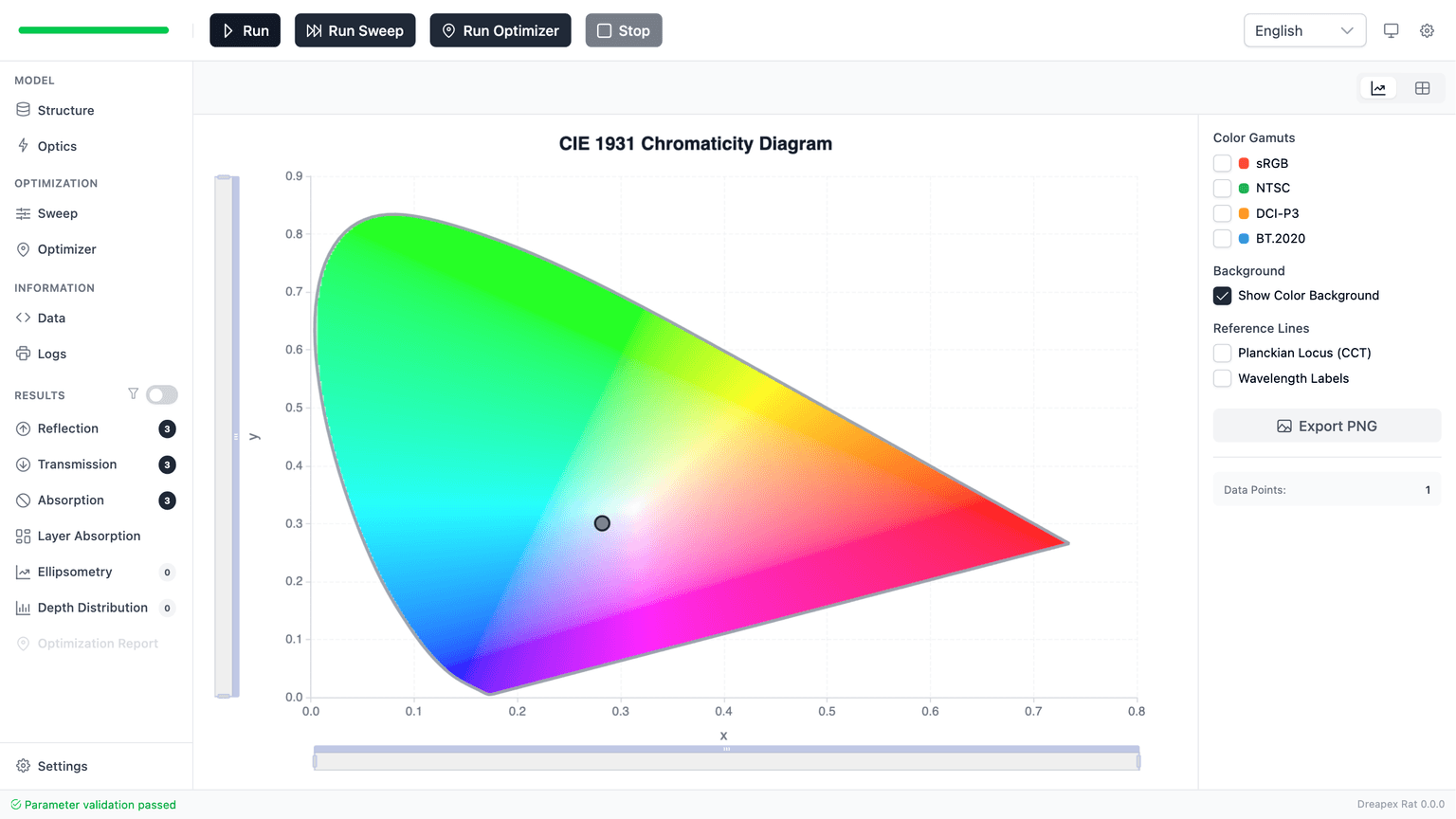
A one-parameter sweep plots one point for each parameter value. Connect Data Points joins them to show the direction of color travel; it changes only the display. Interpret point location together with the parameter value, numeric color difference, and spectrum rather than treating inclusion in a display gamut as a verdict.
Color Data Table
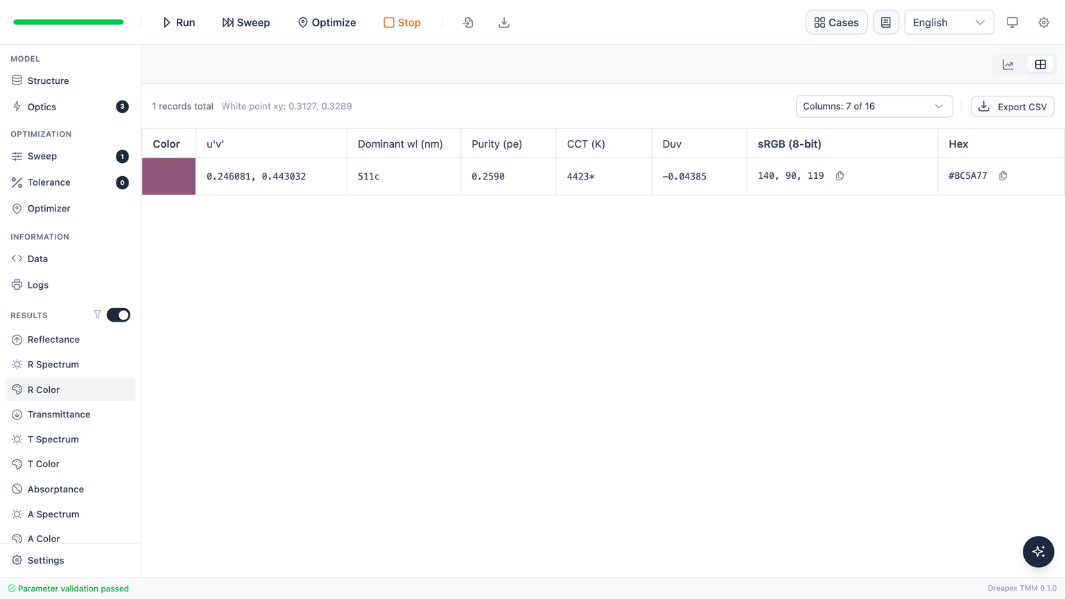
Single runs and sweeps use the same color data table. A single run has one row; a sweep has one row per parameter combination. Columns controls only the on-screen view, while Export CSV always exports the complete audit record.
| Metric group | Fields | Applicable results and use |
|---|---|---|
| Tristimulus and chromaticity | XYZ, xy, xyY | Record spectral integration and chromaticity position; see ISO/CIE 11664-3:2019 for the calculation principles |
| Uniform chromaticity | u′v′ | Compare geometric separation between chromaticity points; see ISO/CIE 11664-5:2024 for the coordinate definition |
| Color descriptors | dominant wavelength, excitation purity, CCT, Duv | Describe hue direction, saturation, and proximity to the white region; available for reflection and transmission |
| Display preview | Linear sRGB, sRGB, sRGB 8-bit, Hex | Screen preview and report colors, not absolute radiometric values |
| Object color | L*a*b*, C*ab, h_ab | Express reflected or transmitted color relative to the incident spectrum; see ISO/CIE 11664-4:2019 for the object-color definition |
| Luminous factor | Y/Yi | Ratio of the sample spectrum's integrated tristimulus to the incident spectrum's tristimulus ; available for reflection, transmission, and absorption |
| Relative to baseline | Delta E00, Delta u′v′ | Color change relative to one baseline row; the former uses the CIEDE2000 formula in ISO/CIE 11664-6:2022 |
Absorbed energy does not reach the observer as a color stimulus. The absorption page therefore omits L*a*b*, C*ab, h_ab, Delta E00, u′v′, dominant wavelength, excitation purity, CCT, Duv, and Delta u′v′. Use absorption Y/Yi to inspect visible-weighted absorption, not as the appearance of a surface.
Color Metrics and Display
XYZ is the common starting point for the color quantities on this page: the result spectrum is integrated with the color-matching functions of the selected CIE standard observer, then converted to xy, u′v′, Lab, and display color. 2° / 10° denote visual-field observers, not incidence angles or accuracy grades; the numbers are also not sampled back from the on-screen swatch. These are simulated colorimetric values, not a physical measurement or compliance report.Incident-Spectrum White Point and Luminous Factor
The L*a*b* white point for reflection and transmission is the incident spectrum used in that run. D65, Illuminant A, and a custom file each produce their own white point. The table header shows its frozen xy, and the CSV records it. Lab values produced under different sources or observers are not directly comparable as one color condition.
Y/Yi is under the actual run conditions: is the tristimulus value of the reflected, transmitted, or absorbed sample spectrum, and is that of the incident spectrum. This dimensionless ratio is displayed as a percentage. It uses the same conditions as commonly reported visible reflection, transmission, or absorption factors only when D65, the 2° observer, normal incidence, and unpolarized light all apply. Under other configurations, interpret it as the ratio for that run.
Baseline Row and Color Differences
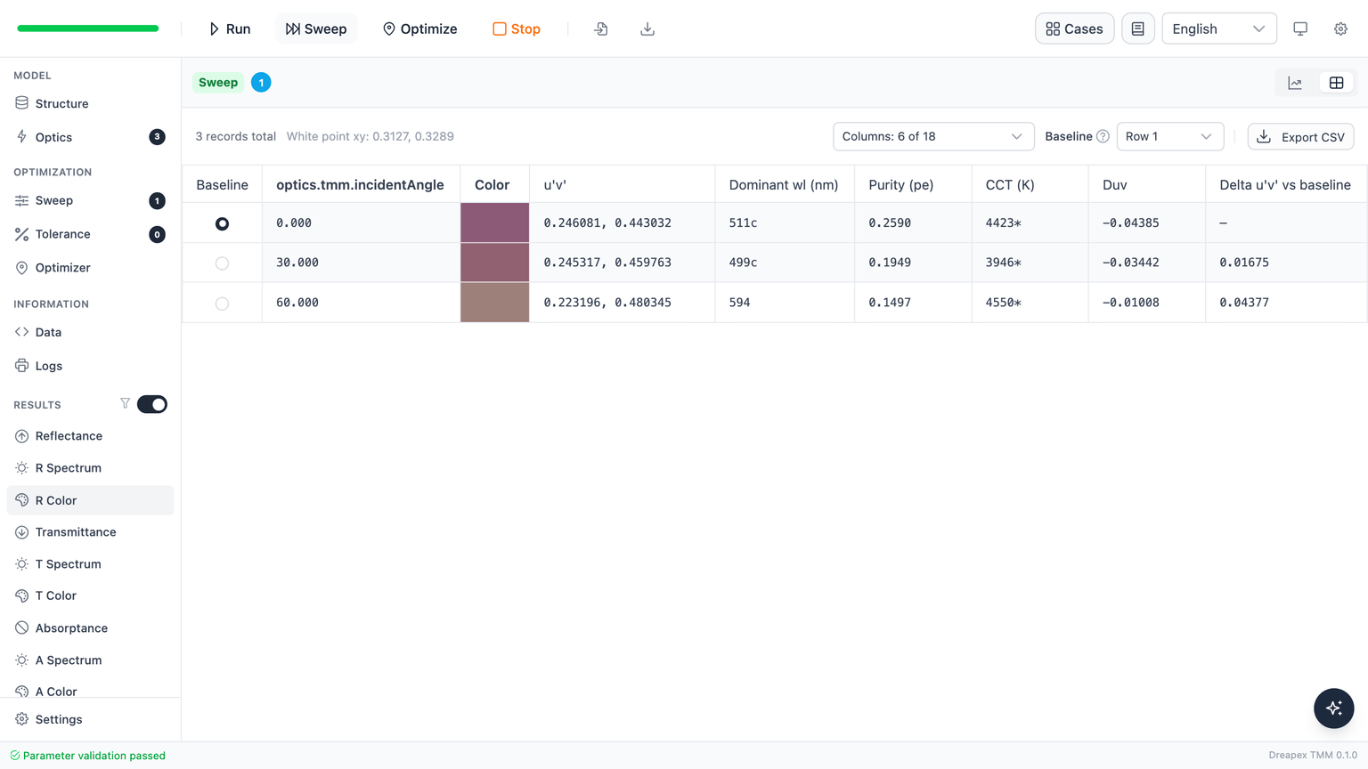
A multi-row table uses the first row as Baseline by default. Select another row with its radio button or the toolbar dropdown. Delta E00 and Delta u′v′ share this baseline, so changing it updates both columns.
Delta E00 uses CIEDE2000. Its lightness, chroma, and hue weighting factors are , , and , respectively, and all three are fixed at 1.
Delta E00 only to compare reflected or transmitted colors under the same illuminant, observer, white point, and calculation conditions. The value has no universal pass/fail threshold; apply the relevant product or customer specification.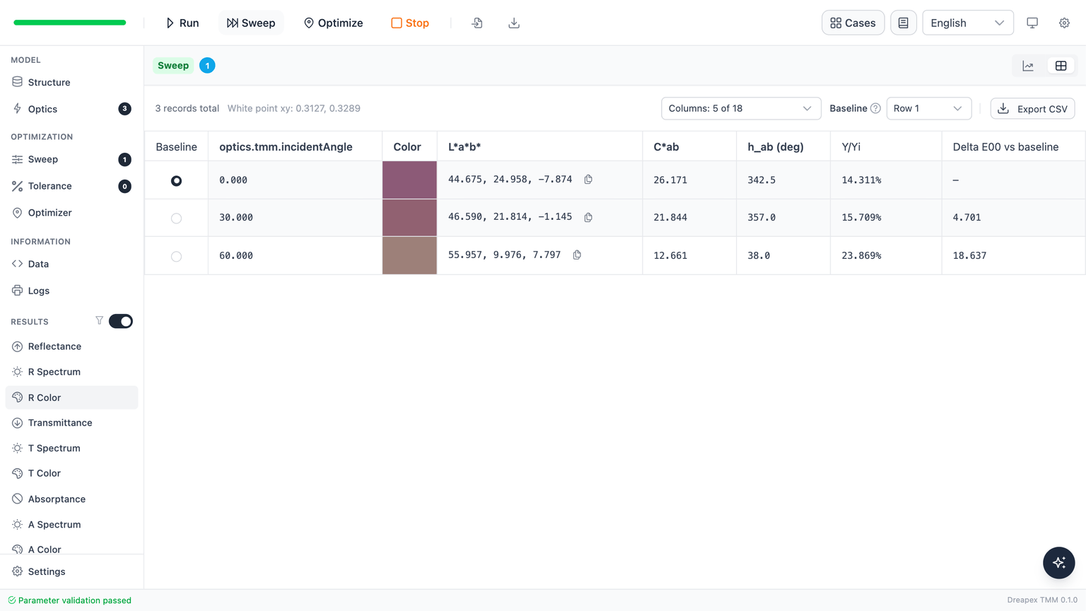
Delta u′v′ is the straight-line distance in the CIE 1976 u′v′ plane. It needs no reference white and has no universal pass/fail threshold; use the product or customer specification. In this example, the reflected color shifts from purple-red at 0° to warm brown at 60°. The 60° row relative to 0° gives Delta u′v′ = 0.04377 and Delta E00 = 18.637, showing a clear change in both hue and lightness at high incidence.
Dominant Wavelength and Excitation Purity
Dominant wavelength describes the perceptual hue direction from an achromatic reference to the current chromaticity point. It is not the peak wavelength of the spectrum. Reflection and transmission use the incident-spectrum white point as their achromatic reference:
- A plain number is the dominant wavelength on the spectral locus, in nanometres.
- A trailing
cdenotes the complementary wavelength for a purple-line color. - When excitation purity
pe < 0.01, the point is too close to the achromatic reference for a stable hue direction, so wavelength is shown as—.
CCT and Duv
CCT and Duv are available only with the 2° observer and use the Ohno (2014) method. CIE TN 013:2022 defines the scope of correlated color temperature near the Planckian locus; use xy, u′v′, and dominant wavelength first for saturated colors:
Duv > 0places chromaticity above the Planckian locus and is typically greener near white;Duv < 0places it below the locus and is typically pinker near white.CCTis shown only when|Duv| <= 0.05; outside that domain,CCTis—whileDuvremains available.- When
0.006 < |Duv| <= 0.05,CCTcarries*. The±0.006reference band is a tool-chosen visual hint, not a binning or compliance verdict.
Run Modes
| Current result | Default view | Table behavior |
|---|---|---|
| Single run | Chromaticity diagram | Toggle to a one-row color table |
One-parameter Sweep | Chromaticity diagram | Toggle between the chart and multi-row table |
Sweep with two or more parameters | Table | Opens directly in the table without a chart toggle |
The chromaticity chart displays at most 2,000 points. Above that limit, use the table and CSV; the data remains available even though all points are not drawn at once.
Next
To screen color change together with R/T/A curves, continue to RTA and Layer Absorption Analysis. To search automatically for a target reflected or transmitted color, continue to Optimizer.